Understanding Joint Dislocation: Causes and Management


Intro
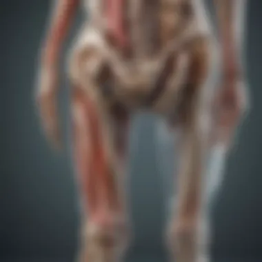
Joint dislocation represents a significant injury within the realm of musculoskeletal health. Understanding the mechanisms behind dislocations, their implications, and effective management strategies is crucial for both medical professionals and laypersons. This injury can occur in various joints, resulting in pain, immobility, and potential long-term complications. Through this analysis, we will explore the definitions, types, causes, symptoms, and treatment protocols related to joint dislocation. An informed perspective can lead to improved awareness and preventative actions for those at risk.
Background and Context
Overview of the Research Topic
Joint dislocation occurs when the bones that form a joint become separated. This often happens due to trauma, such as a fall or collision. The severity of a dislocation can vary based on the joint involved and the extent of the displacement. It's essential to grasp the different types of joint dislocations, as they can require distinct approaches in terms of treatment and rehabilitation.
Historical Significance
Historically, the treatment and understanding of joint dislocations have evolved significantly. Ancient medical texts refer to methods of reducing dislocated joints, often emphasizing the importance of rest and manipulation. Modern medicine has added surgical interventions and advanced rehabilitation techniques to the arsenal of treatment options available today. Knowledge has thus expanded from basic handling of the injury to comprehensive management plans that incorporate both preventative and rehabilitative elements.
“Understanding the historical progression of joint dislocation treatments gives insight into current best practices and why they are effective.”
Key Findings and Discussion
Major Results of the Study
Current studies indicate that both risk factors and management strategies vary widely. Dislocations can be classified into simple and complex, with the latter often involving additional injuries to surrounding structures such as ligaments and tendons. Factors that increase risk include high-impact sports, falls among the elderly, and certain anatomical predispositions.
Detailed Analysis of Findings
The treatment of joint dislocation significantly relies on the type of dislocation, severity, and individual patient factors. Commonly employed methods include:
- Reduction: Realigning the dislocated bones manually or surgically.
- Immobilization: Using splints or casts to prevent movement during the healing process.
- Rehabilitation: Focusing on restoring function, range of motion, and preventing recurrence through targeted exercises.
Evidence suggests that early intervention typically leads to better outcomes and quicker recovery. Understanding the latest research related to joint dislocation enables medical professionals and patients to make informed decisions about treatment options and recovery strategies.
Prelude to Joint Dislocation
Joint dislocation is a critical topic that merits close examination in the medical field. It represents a significant injury that affects not only the physical functionality of joints but also the quality of life of individuals affected. By understanding joint dislocation, practitioners and researchers can enhance their preventive strategies and treatment methodologies. This article aims to demystify the condition and present a thorough overview of various related aspects.
Definition of Joint Dislocation
A joint dislocation occurs when the bones that form a joint become separated. This separation can arise due to various reasons, including trauma, congenital factors, or repetitive stress on the joint. The most commonly dislocated joints are the shoulder, knee, and fingers. Symptoms often include pain, swelling, and an inability to move the affected joint. Immediate medical assessment is essential to confirm the diagnosis and to initiate treatment.
Historical Context and Evolution of Understanding
The medical understanding of joint dislocations has evolved significantly over centuries. In ancient times, treatments were primarily empirical, relying on observation and rudimentary techniques. Early writings in Greek medicine referred to dislocations, but comprehensive understanding emerged only with advances in anatomy and physiology. By the 19th century, with improvements in surgical techniques and imaging, diagnostics became more precise. Today, we recognize joint dislocations not simply as a mechanical issue but closely related to injury mechanisms, anatomical variations, and overall patient health. This historical perspective indicates the progress made and highlights the need for continued research in the field.
"Understanding the historical context of joint dislocation allows for better clinical approaches in current practices."
This journey of understanding showcases how past insights shape present interventions and future innovations.
Anatomy of Joints
Understanding the anatomy of joints is crucial in comprehending joint dislocation and its implications. Each joint serves as a pivotal connection, enabling movement and bearing weight. Grasping the structure and function of joints provides insight into how dislocations occur, the severity of injuries, and approaches for management and rehabilitation. Joint anatomy comprises macroscopic and microscopic components, each contributing to overall joint health and stability.
Basic Joint Structure
Joints can be classified into three main categories: synovial, cartilaginous, and fibrous joints. Synovial joints are the most common, characterized by a space called the synovial cavity filled with synovial fluid. This fluid facilitates movement and reduces friction. The basic structure of a synovial joint includes:
- Articular Cartilage: This smooth layer covers the ends of the bones, providing a cushioning effect to absorb shock.
- Joint Capsule: Encasing the joint, the capsule contains ligaments and synovial fluid, ensuring stability.
- Synovial Membrane: Lining the capsule, this membrane secretes synovial fluid, amping the joint’s lubricating properties.
- Ligaments: These are robust bands of connective tissue that connect bones and offer support during movement.
Injuries to any of these components can lead to dislocation or chronic issues such as arthritis.
Key Components Involved in Joint Function
Several components collaborate to allow smooth and effective joint function:
- Bones: The primary structures that support weight and enable articulation.
- Tendons: Connecting muscles to bones, tendons assist in movement by transmitting force.
- Muscles: Muscle groups surrounding joints provide the necessary strength and stability for movement.
- Nerves: They transmit signals to control and coordinate joint movements.
The interaction of these components is vital. When a joint is displaced, as seen in dislocations, it disrupts this intricate system, leading to pain and loss of function.
In summary, comprehending joint anatomy serves as a foundation for understanding joint dislocation. Recognizing how even minor changes in structure can lead to dislocations underscores the significance of injuries and their management. The implcations ruin not just mobility but also the overall quality of life.
Types of Joint Dislocation
Understanding the various types of joint dislocations is crucial for proper diagnosis and treatment. Each type presents unique characteristics and implications for the affected joint, influencing both clinical approaches and rehabilitation plans. Therefore, knowing these types helps medical professionals make informed decisions and facilitates effective communication with patients regarding their conditions.


Anterior Dislocation
Anterior dislocation occurs when the head of the bone is displaced forward, most commonly seen in the shoulder joint. This type is frequently linked to trauma, such as falls or sports injuries. Patients typically report acute pain and visible deformity at the joint. The prominence of the shoulder may lead to limited mobility, and affected individuals often find any attempt to move excruciating.
Management usually involves immediate immobilization followed by reduction procedures, which can be performed manually in many cases. The rehabilitation post-reduction is crucial, as it helps restore strength and range of motion, emphasizing exercises that strengthen the surrounding muscles.
Posterior Dislocation
In contrast with anterior dislocation, posterior dislocation occurs when the bone shifts backward. Although less common, it often presents in specific circumstances, particularly following seizures or electrical injuries. Symptoms may include severe pain in the shoulder and difficulty with arm movement. Diagnosis typically confirms through imaging like X-rays.
Treatment often requires sedation for reduction due to the severity of the impact. Proper assessment is vital to check for accompanying injuries in the area, as they can complicate recovery and rehabilitation.
Radial and Ulnar Dislocations
Dislocations in the radial and ulnar joints are often seen in the elbow area of young children, specifically from a pull or fall. These dislocations usually result from sudden traction on the arm and can be hard to identify immediately. Symptoms might be subtle, involving discomfort and reluctance to use the arm. Parent observation of unusual behavior can prompt timely medical evaluation.
Reduction of these dislocations often involves simple manipulation techniques. This approach is generally effective, and rehabilitation focuses on restoring function without overstraining the joint.
Other Types of Dislocations
Apart from the common types mentioned, multiple other joint dislocations exist. For instance, dislocations can also happen in the hip, knee, and fingers, each requiring different management strategies depending on specific locations and injury mechanisms. Hip dislocations are particularly urgent, often needing immediate attention due to vascular and neurological risk factors.
Overall, recognizing various types of dislocations assists clinicians in tailoring management strategies to improve recovery times and outcomes. Understanding these distinctions contributes to effective patient education, helping individuals grasp the seriousness of their injuries and understand the necessary steps toward healing.
Causes of Joint Dislocation
Understanding the causes of joint dislocation is crucial for both prevention and effective management of this injury. It allows health professionals to identify at-risk individuals, provide appropriate treatment, and develop rehabilitation strategies. Furthermore, gaining insights into these causes can empower patients to take proactive measures.
Traumatic Injuries
Traumatic injuries represent one of the most common causes of joint dislocation. These injuries usually occur as a result of sudden impact or excessive force to a joint. Common scenarios include accidents, falls, sports injuries, and violent encounters.
The ligaments that support the joints can become overstretched or torn during such events. When they fail to hold the bone in place, dislocation is almost inevitable. For example, dislocations of the shoulder frequently occur in contact sports due to falls and direct hits. Young athletes are particularly vulnerable due to their higher engagement in high-risk activities.
Some signs of dislocation following a traumatic event may include severe pain, visible deformity, and restricted movement. Immediate medical attention is necessary to address traumatic dislocation effectively.
Congenital Factors
Congenital factors can also play a significant role in joint dislocation. These factors refer to conditions present from birth that can weaken the integrity of a joint. For instance, conditions such as Ehlers-Danlos syndrome, Marfan syndrome, or other connective tissue disorders result in hypermobility of the joints, increasing the likelihood of dislocations.
Individuals with these congenital factors may experience recurrent dislocations even with minor activities. A thorough understanding of these underlying conditions is essential for medical professionals to provide appropriate preventive strategies and treatment plans. Genetic predisposition can lead to joint laxity, thus highlighting the importance of family history in the diagnosis and management of joint issues.
Repetitive Stress and Overuse
Repetitive stress and overuse can contribute significantly to joint dislocations, particularly in certain professional and recreational activities. Over time, excessive and repetitive movements can wear down ligaments and tendons surrounding a joint, making it more susceptible to dislocation.
For example, athletes who engage in repetitive motions, such as gymnasts, wrestlers, and tennis players, may find themselves at greater risk. Chronic stress on joints can lead to joint instability, thus increasing the likelihood of dislocation. This kind of injury does not necessarily result from a single event, but rather accumulates over time from continual overexertion.
To prevent dislocations due to overuse, individuals should focus on proper warm-up routines, strength training, and employing correct techniques during activity. Awareness and education on the importance of rest and recovery are vital for maintaining joint health.
It's essential that both athletes and non-athletes understand the mechanics behind joint dislocation to take informed preventive measures.
Through understanding the causes — whether from trauma, congenital issues, or repetitive stress — preventative and management strategies can be better tailored. The insights gained here underscore the significance of individual assessment in developing effective protocols to minimize the occurrence of joint dislocations.
Symptoms and Diagnosis
Understanding the symptoms and diagnostic methods related to joint dislocation is crucial in both acute and long-term management. Identifying these symptoms early can have significant implications for patient outcomes and rehabilitation. By recognizing the signs of joint dislocation, healthcare professionals can quickly implement appropriate interventions, potentially mitigating complications and promoting better recovery.
Common Symptoms of Dislocation
Joint dislocations typically present with several characteristic symptoms. Common indications that a dislocation has occurred include:
- Severe pain: This pain often arises suddenly and is usually intense, making it difficult for the affected individual to move the joint.
- Swelling: Inflammation can occur around the joint, leading to visible swelling.
- Deformity: Dislocated joints may appear misshapen or out of their normal alignment. This can be particularly evident in shoulders and fingers.
- Loss of function: The affected joint often becomes non-functional, resulting in an inability to bear weight or use the limb effectively.
- Numbness or tingling: Depending on the severity of the dislocation, nearby nerves may be affected. A person may experience sensations of tingling, weakness, or numbness.
Recognizing these symptoms promptly can assist in deciding when to seek medical help and enables quicker treatment, which is essential for a favorable prognosis.
Diagnostic Imaging Techniques
Once a dislocation is suspected, effective diagnosis typically involves several imaging techniques. These technologies help healthcare providers visualize the joint and the surrounding structures for precise assessment. Common imaging methods include:
- X-rays: Often the first step in imaging, X-rays can clearly show joint alignment and identify dislocations.
- MRI (Magnetic Resonance Imaging): This technique is effective in assessing soft tissue injuries such as ligaments and tendons surrounding a dislocated joint. An MRI may be used if there is concern for associated injuries.
- CT scans (Computed Tomography): These scans offer detailed cross-sectional images, which can provide further insights into complex fractures or issues not easily visible in X-rays.


Utilizing these diagnostic tools is vital in forming a comprehensive picture of the injury and assisting in deciding the most appropriate treatment protocols.
Clinical Assessment Protocols
A thorough clinical assessment is essential in managing joint dislocations. Healthcare providers often follow a systematic approach to evaluate the extent of the injury. This process includes:
- Patient History: Gathering information about the incident, symptoms, and previous medical history.
- Physical Examination: Assessing the affected joint's appearance, range of motion, and functionality. This examination also includes palpation to check for possible fractures.
- Neurological and Vascular Assessment: Ensuring proper blood flow and nerve function in the affected area is crucial. This includes checking pulses and sensation in the extremities.
A comprehensive clinical assessment facilitates early intervention and can significantly enhance recovery outcomes.
Emerging best practices emphasize the importance of immediate evaluation and subsequent monitoring of dislocated joints to mitigate complications. A detailed understanding of symptoms and diagnosis thus lays the groundwork for effective management and rehabilitation strategies.
Complications Associated with Joint Dislocation
Understanding the complications that arise from joint dislocation is essential for effective management and rehabilitation. Joint dislocation is not simply an isolated injury; it often sets off a cascade of physical and psychological effects that can significantly alter a patient's quality of life. Recognizing these complications can guide healthcare professionals in tailoring treatment plans and inform patients about potential outcomes. This section explores immediate complications, long-term implications, and the psychological impact of dislocation.
Immediate Complications
Immediate complications following a joint dislocation can be severe and often demand prompt medical attention. Common immediate complications include:
- Nerve Damage: Dislocating a joint may stretch or compress nearby nerves, leading to numbness or weakness in the associated limb.
- Vascular Injury: The trauma of dislocation can also affect blood vessels, preventing adequate blood supply to the affected area. This could result in ischemia if not addressed quickly.
- Fractures: In some cases, the force that causes dislocation may also cause bone fractures. This complication requires careful evaluation and treatment.
- Joint Damage: Immediate dislocations may lead to cartilage damage, exacerbating the injury and complicating recovery.
These complications may not always be apparent immediately and might require diagnostic imaging techniques to assess. Timely diagnosis and management are critical. Failure to address these complications could lead to more severe consequences in the future.
Long-term Implications
Long-term implications of joint dislocation can lead to chronic health issues if not managed correctly. Some potential long-term complications include:
- Joint Instability: Recurrent dislocations can lead to chronic instability in the joint. This often necessitates surgical intervention.
- Arthritis: Cartilage damage from the initial injury can predispose individuals to osteoarthritis, leading to joint pain and reduced mobility over time.
- Loss of Range of Motion: Scar tissue formation can limit a patient’s ability to recover full range of motion in the affected joint.
- Osteonecrosis: In severe cases, interrupted blood flow can lead to bone death, significantly affecting joint function.
Patients should be educated on the possibility of these long-term complications. Consistent follow-up care and appropriate rehabilitation are vital for minimizing these risks.
Psychological Impact of Dislocation
The psychological impact of joint dislocation should not be overlooked. An injury like this can induce various mental health issues, commonly manifesting as:
- Anxiety: Fear of re-injury can cause anxiety, inhibiting participation in physical activities.
- Depression: Chronic pain and the inability to engage in regular daily activities due to dislocation can lead to feelings of helplessness and depression.
- Post-Traumatic Stress Disorder (PTSD): For some individuals, the traumatic nature of the injury can lead to PTSD, requiring specialized treatment.
It is crucial for healthcare professionals to screen patients for these psychological effects and incorporate mental health support into the rehabilitation process. This may involve referrals to psychologists or counselors specializing in sports or injury-related trauma.
Key Takeaway: Understanding complications associated with joint dislocation enhances management strategies, promoting better long-term outcomes for patients.
Management of Joint Dislocation
The management of joint dislocation is a crucial aspect that aims to restore the joint's normal position while minimizing complications and ensuring optimal recovery. Timely and effective treatment can significantly influence the outcomes. Immediate attention can prevent further injury, reduce pain, and enable a smoother recovery process. An understanding of management strategies helps both medical professionals and individuals involved in sports or physical activities to respond appropriately when dislocations occur.
Immediate First Aid Measures
Immediate first aid measures for a dislocated joint are essential in reducing further damage and pain. Recognizing the signs of a dislocation can prompt timely action. The following steps can guide initial responses:
- Do not try to relocate the joint on your own. This could exacerbate the injury.
- Immobilize the joint carefully. Using a makeshift splint or supports can prevent movement and additional stress.
- Apply ice packs to the affected area. Cold therapy aids in reducing swelling and pain.
- Keep the person calm. Anxiety can increase tension, which may worsen the situation.
Always seek professional medical assistance as soon as possible. Effective first aid can minimize complications and help preserve joint function.
Reduction Procedures
Once medical help is sought, reduction procedures are typically employed by healthcare professionals. This process involves maneuvering the dislocated joint back into its normal anatomical position. There are several techniques used:
- Closed reduction: This method requires no surgical incisions. The healthcare provider uses specific movements to coax the joint back into its socket.
- Open reduction: If closed reduction is unsuccessful or if there are associated fractures, surgical intervention may be necessary to realign the joint properly.
The choice between these methods will depend on the type of dislocation, accompanying injuries, and the patient's overall health. It is important to conduct reduction procedures in a safe, controlled environment to prevent complications such as nerve damage.
Surgical Intervention
In certain situations, surgical intervention may be the best option for managing joint dislocation. This is usually the case when:
- The dislocation is complicated by associated fractures.
- There are long-term issues, like recurrent dislocations, that standard reduction cannot address.
- Significant ligament damage is present that requires repair.
The surgery aims to restore the joint's stability and function. Recovery from surgical intervention typically involves a longer rehabilitation period. Patients can expect to follow a structured physical therapy program that focuses on restoring strength and range of motion. Addressing joint dislocation promptly through these management strategies is vital for both recovery and preventing future dislocations.
Rehabilitation Post-Dislocation


Rehabilitation post-dislocation is a crucial stage in the recovery process. Once a joint dislocation has been treated, whether through nonsurgical methods or surgical intervention, a structured rehabilitation program becomes essential. The emphasis on rehabilitation lies in restoring function, reducing the risk of future dislocations, and enhancing overall joint stability. A comprehensive approach includes physical therapy, education, and gradual return to normal activities.
Initial Recovery Phase
The initial recovery phase is vital for laying the foundation for rehabilitation. This phase usually begins soon after the joint has been relocated. Emphasis is placed on protecting the injured area to prevent further damage. Immediate tasks involve managing pain and swelling through methods such as ice therapy and appropriate elevation.
During this time, passive range-of-motion exercises may be introduced, focusing on keeping the surrounding muscles engaged without stressing the joint. It is essential to connect to a healthcare provider, who can guide the recovery process with personalized advice. Key considerations during this phase include:
- Rest the joint to allow healing
- Use ice and compression to manage swelling
- Consult a physician to assess the condition
Physical Therapy Regimens
Physical therapy regimens play a significant role in recovery from joint dislocation. Once the initial pain and swelling diminish, a structured program with a physical therapist can help restore strength and flexibility to the affected joint. These regimens typically progress in four main areas: flexibility, strength building, functional skills, and overall fitness.
- Flexibility: Stretching exercises are essential to regain range of motion. This may include gentle stretches guided by a therapist.
- Strengthening: Targeted exercises focus on muscles around the joint to ensure stability and support. Resistance bands or weights might be introduced gradually.
- Functional Skills: Relearning specific movements or activities adapted to the individual's needs is important for a full return to activities.
- Overall Fitness: Maintaining general fitness through low-impact aerobic exercises can aid recovery and enhance well-being.
Careful monitoring of progress is crucial, as pushing too hard or too fast can lead to setbacks.
Return to Activity Guidelines
The return to activity guidelines are critical for a safe transition back to regular routines and sports. It is essential to proceed with caution and wait until the healthcare providers recommend it. Patient readiness can vary significantly depending on factors like the type of dislocation, the treatment received, and individual healing rates.
A gradual approach is recommended:
- Start with light activities: Engage in low intensity exercises to gauge comfort levels.
- Listen to your body: Pay attention to signs of discomfort or pain and adjust activities accordingly.
- Progress to moderate activities: Slowly reintroduce more demanding physical tasks as strength and confidence improve.
- Consult professionals: Ongoing communication with therapists or physicians ensures that the return to activity is safe and effective.
Prevention of Joint Dislocation
Preventing joint dislocation is crucial for individuals, especially athletes and those engaged in physically demanding activities. Dislocations can lead to serious injury and prolonged recovery times. By focusing on proper prevention strategies, it is possible to significantly reduce the likelihood of dislocations occurring. There are specific elements involved in prevention, including strengthening exercises, proper techniques in sports, and the use of protective gear.
Strengthening Exercises
Strengthening exercises play a key role in joint stability. The stronger the muscles surrounding the joint, the better they can support and protect it. For instance, exercises targeting the shoulder, knee, and ankle joints can help build the necessary muscular support.
- Focus on targeted workouts that enhance stability.
- Incorporate resistance training to fortify ligaments.
- Balance exercises can improve proprioception, which is vital for maintaining joint alignment.
For example, rotator cuff exercises for shoulder strength can be helpful. Lower body exercises, like squats and lunges, are effective for knee joint protection. Consistency in doing these exercises enhances resilience, thus minimizing the risk of dislocation.
Proper Techniques in Sports
Proper techniques during sports activities are essential for avoiding injuries, including dislocations. Athletes must receive training on movement patterns that mitigate the risk of joint injury. Simple adjustments can make a significant difference in joint safety.
- Coaches should emphasize correct body mechanics.
- Athletes must learn ways to fall or land safely.
- Regular drills can reinforce safe practices.
For instance, in contact sports like football, teaching players how to tackle appropriately can prevent shoulder dislocations. In gymnastics or diving, correct landing techniques are vital for wrist and shoulder stabilization. Understanding and implementing these techniques dramatically lowers the chances of dislocations occurring during high-risk activities.
Use of Protective Gear
The use of protective gear serves as an additional line of defense against joint dislocations. Wearing appropriate equipment can absorb impact and reduce the stress placed on joints during activities.
- Items like knee braces, ankle supports, and shoulder pads can safeguard vulnerable areas.
- For specific sports, it is important to use sport-specific protective gear.
For example, skiers should wear knee guards, while hockey players benefit from knee and shoulder pads. It is also crucial to ensure that the equipment fits properly. Wearing ill-fitting gear may lead to instability rather than protection, increasing dislocation risk. Properly chosen and fitted protective gear is essential in the prevention strategy against joint dislocation.
Investing time and effort in prevention not only protects the individual but also promotes long-term athletic and physical health.
The End
The conclusion serves a pivotal role in synthesizing the information presented throughout the article on joint dislocation. It is the moment where insights blend into a cohesive understanding. Throughout the article, we have navigated the intricate landscapes of joint dislocation, from its definition to its management and prevention. Now, it is essential to frame the significance of these discussions.
One key element in the conclusion is the reinforcement of the importance of awareness. Joint dislocations, while common, can lead to severe complications if not properly addressed. Understanding the complexities—from symptom recognition to the appropriate treatment methods—ensures that individuals can respond effectively when faced with this injury.
Furthermore, the benefits of this compilation extend to educating not just healthcare professionals but also the general public. When people are informed about the risks and recovery processes associated with dislocations, they are more likely to engage in preventive measures like strengthening exercises and the use of protective gear.
Consideration of future developments in research is also highlighted within the conclusion. The evolving nature of medicine means that new insights into joint dislocation will undoubtedly emerge. Staying abreast of these advancements can promote better patient outcomes and improved rehabilitation processes.
In summary, the conclusion encapsulates the heart of the article. It emphasizes ongoing education, the necessity of prevention strategies, and the importance of research to foster a well-rounded comprehension of joint dislocation. By addressing these areas, we not only increase awareness but also empower individuals to handle potential dislocations with greater confidence and knowledge.
Summary of Key Points
- Definition and Types: Joint dislocation involves the displacement of bones at a joint, with various types such as anterior and posterior dislocations being prevalent.
- Causes: Dislocations can arise from traumatic injuries, congenital conditions, or repetitive stress.
- Symptoms and Diagnosis: Recognizing symptoms such as pain and immobility is crucial for timely diagnosis and treatment.
- Management: Effective management includes immediate first aid, reduction procedures, and possible surgical interventions in severe cases.
- Rehabilitation and Prevention: Post-dislocation rehabilitation is fundamental to restore function and prevent recurrence through targeted exercises and preventive techniques.
Future Directions in Research
Looking ahead, the research on joint dislocation is likely to expand in various critical areas. Some key directions are:
- Improving Diagnostic Techniques: Innovations in imaging technology may lead to more accurate and timely diagnoses.
- Biological Healing Processes: Understanding the biological processes during recovery could inform better rehabilitation practices.
- Long-term Impact Studies: More extensive studies on the long-term implications of dislocation may reveal connections to chronic joint issues.
- Prevention Tactics: Research aimed at developing new prevention strategies tailored for specific populations, such as athletes or individuals in high-risk professions, can enhance outcomes.
By pursuing these avenues, the medical community can bolster the understanding of joint dislocation and improve treatment protocols.


