Exosome Isolation from Tissue: Methods and Applications

Intro
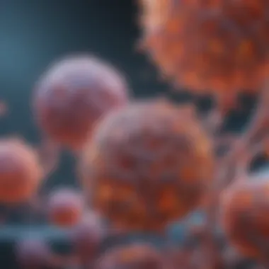
Exosomes, those minuscule vesicles with a big impact, are becoming a focal point in biomedical research. Their role as messengers in cellular communication opens a Pandora's box of opportunities in diagnostics and therapeutics. Understanding how to isolate these treasures from tissue samples effectively is crucial for advancements in the field. But wading through the various isolation techniques, each with its own set of challenges, is like navigating a minefield.
This article will untangle the complexities surrounding exosome isolation from tissue, starting with the essential background that led us to this point. We'll explore the methods researchers employ, discuss potential obstacles, and delve into the practical applications in clinical settings. By the end, you will have a robust understanding of the intricate dance between tissue types and exosome isolation, as well as insight into current and future research directions.
Background and Context
Overview of the Research Topic
When we talk about exosomes, we're referring to the small extracellular vesicles that pack a punch in cellular communication. Their size usually ranges from 30 to 150 nanometers, yet these little guys hold a wealth of information. They transport proteins, lipids, and RNA, making them a critical component in the dialogue between cells. As researchers dive deeper into their functions, the need to extract them from various tissues becomes paramount.
Historical Significance
The study of exosomes isn't brand new; it has roots in the early 1980s when scientists first identified them. Initially considered mere cellular debris, exosomes were largely overlooked. However, fast forward to the past decade, and these vesicles have transformed into diagnostic tools and therapeutic agents. Their potential in liquid biopsies and targeted drug delivery systems has sparked a wave of interest across multiple disciplines, making the meticulous art of exosome isolation an essential focus.
Key Findings and Discussion
Major Results of the Study
In analyzing the various isolation techniques available today, several key findings have emerged. Researchers have employed methods ranging from ultrafiltration to immunoaffinity capture, each boasting unique advantages and limitations. Recent studies indicate that the integrity and yield of exosome isolation can vary significantly based on the chosen method and the type of tissue being examined.
Detailed Analysis of Findings
- Ultracentrifugation is often lauded for its effectiveness, making it a common first choice. However, it’s not without its drawbacks, notably the time it consumes and the technician's expertise required.
- Precipitation-based methods offer speed and simplicity, but they may compromise exosome purity due to the presence of contaminants.
- Size-exclusion chromatography has emerged as a promising alternative, providing a balance between purity and yield, though it can still struggle with certain tissue types.
- Immunoaffinity capture, while highly specific, can sometimes yield low quantities of exosomes, especially from samples that are inherently low in these particles.
The diversity of isolation techniques reflects the complex biology of exosomes themselves. Adjustments must often be made based on sample type, the purpose of isolation, and downstream applications.
Challenges Faced
Aside from the technical hurdles, researchers also grapple with the biological implications of isolating exosomes. Tissue type can markedly influence the yield, functionality, and physicochemical properties of exosomes. Moreover, standardized protocols are still in short supply, which hampers reproducibility across studies. With the push for personalized medicine, addressing these challenges becomes even more vital.
Conclusively, the field of exosome isolation from tissue is vibrant and evolving, marked by innovative techniques and burgeoning applications. As we venture further into detailed methodologies in the upcoming sections, the overarching theme remains clear: the interplay between isolation methods and biological contexts holds the key to unlocking the full potential of exosomes in diagnostics and therapeutics.
Prologue to Exosomes
Exosomes have become a sizzling topic within the realm of biomedical research, serving as crucial mediators in various physiological and pathological processes. Understanding exosomes—tiny vesicles secreted by cells—holds immense value in the broader context of molecular biology, therapeutics, and diagnostics. In the landscape of cell communication, exosomes are tiny parcels packed with information that cells delicately exchange. Thus, expanding our understanding of exosomes is not just a niche interest; it's pivotal for numerous fields, including regenerative medicine, cancer therapy, and neurobiology.
Defining Exosomes
Exosomes are membrane-enclosed vesicles that range from 30 to 150 nanometers in diameter. They differentiate themselves from other vesicles by their distinct biogenesis pathways. Exosomes are formed within the endosomal system, particularly as intraluminal vesicles of multivesicular bodies. Released into the extracellular space upon fusion with the plasma membrane, these exosomes carry proteins, lipids, and nucleic acids, acting as molecular messengers. Given their diverse cargo, they can influence neighboring cells or even act at a distance.
Some key feature include:
- Size: Typically 30 to 150 nm, smaller than many cellular components
- Biological origin: Derived from the endosomal system, creating a discrete environment for cargo sorting
- Cargo: Contains proteins like tetraspanins, lipids, and various RNA types, including mRNA and miRNA.
Understanding their structure and the molecular makeup is vital as we examine their roles in health and disease.
Biological Significance
The biological relevance of exosomes extends beyond simple cellular debris. They play a major role in numerous physiological and pathological functions. The scope of their impact can be broadly categorized into several domains:
- Intercellular Communication: Exosomes carry signals that can modulate the behavior of recipient cells. This function is especially crucial in cancer, where they may transfer oncogenic factors that help in tumor progression.
- Immune Modulation: Exosomes influence the immune response, assisting in antigen presentation and mediating immune tolerance. Some studies suggest they can act in such a way as to suppress immune responses, which may be advantageous for tumor cells.
- Biosignaling: The cargo contained within exosomes often reflects the physiological state of the secreting cell, making them potential biomarkers for disease detection and monitoring.
"Exosomes exhibit an ability to influence their surroundings in ways both profound and subtle—understanding these vesicles is tantamount to grasping the complexities of cell communication."
These aspects make the study of exosomes imperative, not only for understanding fundamental biological processes but also for practical applications in diagnostics and therapies. By delving into their roles and functions within various environments, we can harness the insights gleaned for innovative strategies in disease management.
In a nutshell, exosomes represent more than mere cellular refuse; they are pivotal players with profound implications in various biological contexts and emerging fields. As we transition to discussing the importance of isolating these exosomes, it becomes clear that their implications for diagnostics and therapeutics cannot be overstated.
Importance of Exosome Isolation
Exosome isolation plays a pivotal role in advancing biomedical research, especially in the realms of disease diagnostics and therapeutics. These nanoparticles, secreted by various cells, act as messengers carrying crucial biomolecules that inform not only on normal cellular functions but also on disease states. Understanding the importance of isolating exosomes enhances their applications in clinical settings, making it a vital topic.
Role in Disease Diagnostics
The significance of exosome isolation in disease diagnostics cannot be overstated. As carriers of specific proteins, lipids, and RNA, exosomes are a mirror reflecting the physiological state of their parent cells. For instance, cancer cells release exosomes rich in tumor-associated markers. When exosomes are effectively isolated from biological fluids like blood or urine, they enable the identification of these makers, which could herald the development of non-invasive diagnostic tools.
"Isolating the right exosomes could lead to breakthroughs in early cancer detection, allowing for timely intervention."
Such potential has been evidenced in various studies showing that protein and RNA profiles from exosomes can assist in differentiating between types of cancers, even at early stages. Furthermore, because exosomes can be derived from tissues affected by various diseases, they hold the promise of offering a comprehensive view of disease mechanisms. They could also play a role in predicting disease progression, giving practitioners a solid basis for tailored treatment plans.
Therapeutic Potential
The therapeutic utility of isolated exosomes extends beyond their role as biomarkers. They possess intrinsic properties that make them candidates for novel therapeutic strategies. For example, exosomes can carry therapeutic agents such as small interfering RNA (siRNA) and proteins, enabling targeted delivery to specific cells. This targeted approach can minimize side effects often associated with traditional therapies.
Moreover, researchers are investigating the potential of exosomes in regenerative medicine. Isolated exosomes from stem cells have demonstrated capabilities to promote tissue repair and regeneration, thereby paving the way for innovative treatments in fields like orthopedics and cardiology.
In essence, as we unravel the complexities of exosomes, their isolation stands out as a critical step toward harnessing their diagnostic and therapeutic prowess. The concerted efforts in optimizing isolation techniques can not only enhance research but also translate discoveries into tangible clinical applications.
Sources of Exosomes in Tissue
Understanding the sources of exosomes in tissue is paramount for researchers aiming to unravel the complexities of cell communication and the role these nano-sized vesicles play in various physiological and pathological conditions. Exosomes derived from different tissues can carry distinct molecular signatures, fundamentally shaping their potential applications in diagnostics and therapeutics. Grasping these variances can inform choices made in exosome isolation methodologies and ultimately impact research outcomes and clinical strategies.
Tissue Types and Exosome Profiles
Exosomes are not one-size-fits-all; their makeup varies considerably across different tissue types. For instance, exosomes derived from cancerous tissues often reflect the altered metabolic states of tumor cells, featuring unique proteins and RNAs that differ from those in normal tissues. This differentiation can be crucial when attempting to develop cancer biomarkers.
Another good example is the role of exosomes from immune cells, which are involved in orchestrating immune responses and can provide insights into inflammatory diseases. They sprinkle their cargo with critical molecules that can both activate and silence various pathways.
Consider the following points about exosome profiles from assorted tissues:
- Cancer Tissues: These exosomes can carry oncogenic mutations, serving as vehicles for anti-tumor therapies or enhancing tumor spread.
- Neural Tissues: The exosomal content may include neurotrophic factors, playing a crucial part in neuron repair and signaling, particularly in degenerative conditions.
- Cardiac Tissues: Heart-derived exosomes can carry microRNAs linked with cardiovascular health, useful for predicting myocardial infarction risks.
Such distinct profiles illustrate that isolating exosomes from the correct tissue is essential for target-specific applications. By aligning the source of exosomes with the intended clinical or research use, we can tailor strategies for their effective utilization.
Impact of Tissue Microenvironment
The microenvironment surrounding a tissue serves as a significant influencer of exosome composition and function. Factors within this microenvironment—including nutrient availability, stress signals, and cellular interactions—can modulate exosomal cargo. For instance, in a hypoxic environment, cancer cells might release exosomes rich in angiogenic factors that promote blood vessel growth to support tumor survival.
Moreover, this context can affect the stability and functionality of exosomes outside their originating tissue. Environments rich in reactive oxygen species may alter exosomal integrity, while inflammatory conditions could affect exosome release rates. These points highlight why researchers must consider tissue microenvironments, as failing to account for them may lead to misguided conclusions about exosome pathology or functionality.
Isolation Techniques for Exosomes
Exosome isolation is a fundamental step that carries significant weight in research and therapeutic applications. From diagnostics to targeted therapies, the methods used to extract these nanoscale vesicles can influence the quality and efficacy of downstream experiments. Different isolation techniques are not just varied in methodology, but they also come with unique strengths and weaknesses that researchers must carefully consider. The selection of a particular isolation method can affect exosome yield, purity, and overall functionality, making it crucial to choose wisely.
Ultracentrifugation
Principle of Ultracentrifugation
Ultracentrifugation is widely recognized for its ability to separate exosomes based on their density. It relies on the application of high centrifugal forces, allowing heavier particles to sediment faster than lighter ones. This method proves to be effective in isolating a broad range of exosomes, thus making it a trusted choice for those in the field.
A key characteristic of ultracentrifugation is its throughput capacity. It can process multiple samples concurrently, which is beneficial for studies requiring large datasets. However, it requires specialized equipment and trained personnel, presenting a barrier for some laboratories. The uniqueness of this method lies in its efficiency at isolating high-purity exosomes, though it may also co-purify other unwanted cellular debris.
Protocols and Variations
Protocols for ultracentrifugation can vary significantly depending on the specific objectives of the isolation. Variations include adjustments in time, centrifugal force, and temperature to optimize exosome yield and purity. Many researchers have developed customized protocols based on their unique sample types or desired outcomes, demonstrating the flexibility of this technique.
The benefit of these variations is that they allow for tailoring the isolation process, which can enhance the effectiveness of downstream applications. However, inconsistent methodologies can lead to reproducibility issues in multi-laboratory studies. Standardizing processes across different labs remains a challenge for this approach.
Advantages and Disadvantages
The major advantage of ultracentrifugation is its proven effectiveness in isolating exosomes with high yield and purity, making it a reputable method. Nevertheless, this technique also has its downsides. The need for expensive equipment and extended processing times can be seen as a hindrance, particularly for smaller labs or those just starting in exosome research.
Ultracentrifugation remains a cornerstone in exosome isolation despite its logistical challenges, offering unrivaled purity when executed properly.
Filtration Methods
Types of Filters
Filtration methods offer an alternative to ultracentrifugation, using various filter types such as membrane filters and microfilters. These filters differ in pore size, allowing for selective separation of exosomes from larger contaminants, such as cells or cell debris. A defining feature of filtration methods is their ease of use, enabling quicker processing times and lower costs.
Using filtration is beneficial for labs looking for efficient processing without the extensive equipment required for centrifugation. However, low-pore-sized filters may lead to clogging and require careful monitoring to maintain optimal flow rates.
Application in Exosome Isolation
The application of filtration methods can simplify the isolation of exosomes and is especially effective when dealing with high volumes of fluid. This technique can be utilized alongside other isolation methods, in a complimentary fashion, to enhance purity and efficiency further. Certain filters can also reduce the risk of contamination, a common concern in exosome research.
However, limitations arise as filtration methods don't sufficiently isolate very small exosomes or handle complex biological samples, which can include numerous interfering substances. Thus, understanding the specific needs of a study is vital when considering this approach.
Limitations
Despite their advantages, filtration methods come with drawbacks. One significant limitation is the inconsistency in the size and shape of exosomes, which can lead to variability in purity and yield. Additionally, their inability to fully separate exosomes from other small vesicles can affect downstream applications and diagnostics, making it crucial to choose the right tool for the job.
Precipitation Techniques
Commercial Reagents
Precipitation techniques leverage various commercial reagents designed for isolating exosomes through the aggregation of vesicles. The ease of use and the relatively quick process are distinct features that turn precipitation into an appealing choice. Many commercial kits are available that provide detailed protocols and consistent results, a crucial factor for labs lacking resources or experience with more complex isolation techniques.
However, the wide variety of reagents can lead to differences in outcomes. The cost associated with these kits can also be a deterrent, especially for startups or smaller labs operating on limited budgets.
Effectiveness and Considerations
The effectiveness of precipitation methods can vary based on the type of reagents used and the biofluids from which exosomes are isolated. They are generally considered effective for isolating modest amounts of exosomes, yet they may not provide the same level of purity as ultracentrifugation. Considerations should involve the specific application of isolated exosomes; for example, clinical diagnostic uses may necessitate higher purity than what precipitation methods can achieve.
Comparison with Other Methods
When comparing precipitation techniques with ultracentrifugation and filtration, they often lag in yield and purity. While they are convenient and straightforward to implement, they may fall short in fulfilling the stringent purity requirements demanded by certain research applications. Therefore, researchers must weigh the pros and cons carefully when selecting their isolation strategy.
Affinity-based Techniques
Targeting Specific Markers
Affinity-based techniques utilize the principle of specific binding to target markers on the surface of exosomes for isolation. This method enhances selectivity, allowing for the isolation of exosomes that are of particular interest based on their surface proteins. This targeting feature can substantially improve the quality of the isolated exosomes, particularly in complex samples.
Such specificity ensures that downstream applications are only using exosomes that meet the necessary criteria, which can be crucial in studies investigating particular diseases or biological pathways. However, identifying the right markers and their antibodies can be time-consuming and require extensive research.
Prospects for Customized Isolation
With the advent of personalized medicine, the prospects for customized isolation using affinity-based methods are particularly promising. Researchers can tailor the isolation protocols to target exosomes from specific diseases or physiological conditions, enhancing the relevance of their findings. Nonetheless, developing these specific methods may result in additional costs and longer study timelines.
Research Applications
The applications of affinity-based isolation in research are significant. By honing in on specific exosome populations, researchers can gain insights that would be missed with broader techniques. This targeted approach, however, does require careful validation to confirm that the isolated exosomes truly represent the intended population.
Microfluidic Devices
Emerging Technologies
Microfluidic devices represent a new frontier in exosome isolation that takes advantage of small-scale handling of fluids. These technologies offer a highly controlled environment for separation processes, allowing for faster and more efficient isolations. The ability to perform multiple tests or isolations simultaneously makes these devices particularly attractive for high-throughput applications.
However, they require specialized knowledge and equipment, which may not be readily available in all labs. Furthermore, scaling up from lab prototypes to full-fledged clinical devices poses another significant challenge, as does ensuring consistency across different device batches.
Precision and Efficiency
One of the hallmarks of microfluidic technology is precision. By manipulating tiny fluid volumes, it allows for more efficient recoveries with less waste of biological samples. This level of precision also reduces the risk of contamination, a significant benefit in sensitive experiments.
While their advantages are clear, the complexity of their design and operation may limit their accessibility for some researchers, necessitating collaboration with engineers or specialized developers.
Future Directions
As technology evolves, microfluidic devices are poised for greater integration into exosome research. Future directions may include enhanced miniaturization, automation, and the development of user-friendly interfaces. This evolution could make exosome isolation accessible to a broader range of labs, fostering advancements in diagnostics and therapeutics.
The journey of isolating exosomes from tissues is not just a one-size-fits-all process; it requires a keen understanding of the available techniques and their implications on the quality and performance of the exosomes for future applications.
Challenges in Exosome Isolation
Exosome isolation, despite its significance, is not without its hurdles. The importance of addressing these challenges is paramount, as they can directly impact the quality and reliability of the results obtained in both diagnostics and therapeutic contexts. Identifying the specific elements and considerations surrounding the isolation process can enhance reproducibility and utility across various scientific studies.
Contamination Issues
Contamination during exosome isolation is a prevalent concern in research. When tissues are processed, several contaminants such as proteins, lipids, and even nucleic acids can co-precipitate alongside exosomes. This can create a jumble of unwanted materials, effectively muddying the waters and complicating downstream analyses. It's like trying to find a needle in a haystack; if the haystack is too large and dense with foreign material, that needle (the pure exosome) can easily be lost.
To mitigate contamination, researchers often employ rigorous centrifugation or filtration steps. However, some contaminants — especially those with similar density or size — can escape these methods, leading to a low yield of pure exosomes. Monitoring the process carefully can be essential, as is validating the purity using biophysical techniques such as nanoparticle tracking analysis or electron microscopy. By doing so, one can not only ensure the quality of isolated exosomes but also foster confidence in subsequent analyses and applications.
Variability in Exosome Yield
The variability in exosome yields from tissue samples poses another challenge. Every tissue type can produce exosomes but the quantity and quality vary significantly. For example, the exosomes derived from tumors can differ from those obtained from healthy tissues in terms of size, composition, and even functional properties. This variability is influenced by such factors as the physiological state of the tissue, the isolation method employed, and the duration since tissue collection.
Moreover, not all methods guarantee the same yield. Some researchers might find that ultracentrifugation yields a higher amount of exosomes from one sample yet produces a lower yield from another. Variations in handling, time delays, and even environmental conditions can compound these differences, making comparability between studies arduous. Recognizing these quirks is crucial for ensuring robust applications in diagnostics and treatments. Being aware of these nuances allows researchers to adopt suitable isolation techniques and standard operating procedures that complement the specific tissue type.
Standardization of Protocols
The lack of standardized protocols for exosome isolation remains a significant barrier to research. As the field evolves, different laboratories adopt various methods, often leading to conflicting results. This lack of uniformity can hinder collaborative research efforts and make it difficult for findings to be reproduced. Organizations like the International Society for Extracellular Vesicles are actively striving to define consolidated guidelines and best practices; however, this is still a work-in-progress.
To foster consistency, researchers are encouraged to document their methods meticulously and share their experiences. Standardized protocols, such as specific centrifugation speeds or reagent concentrations, can lead to more reliable results across the board. In doing so, a common language can emerge within the scientific community, bolstering the credibility of findings and advancing the field in a meaningful way.
"Standardization in exosome isolation can pave the way for enhanced reproducibility and reliability, inspiring trust in the diagnostic and therapeutic applications of these tiny, impactful vesicles."
In summary, while significant hurdles exist in exosome isolation, addressing these challenges offers the path forward. By confronting contamination, yield variability, and the lack of standard protocols, researchers can work towards creating a more predictable and impactful future for exosome-related studies, applications, and innovations.
Applications of Isolated Exosomes
Isolated exosomes have emerged as pivotal agents in various fields, particularly in diagnostics and therapeutics. Their natural origins, coupled with their unique properties, make them promising tools in medical applications. As exosomes are rich in proteins, lipids, and nucleic acids, they serve as informative biomarkers and effective carriers for therapeutic agents. This section delves deeper into the specific roles that isolated exosomes play in biomarker discovery, diagnostics, and therapeutic development.
Biomarkers and Diagnostics
The potential of exosomes as biomarkers is nothing short of revolutionary. They carry information reflective of their cell of origin, thus serving as surrogates for disease states. For instance, cancer cells release exosomes into the circulation, which can be analyzed for early detection. The composition of these exosomes can provide insights into the tumor microenvironment, enabling the identification of specific markers associated with particular cancers.
Key benefits of using exosomes for diagnostics include:
- Non-Invasive Sampling: Analyzing exosomes from blood, urine, or saliva is considerably less invasive than obtaining biopsies.
- Wide Range of Applications: Exosomes can be used to monitor various diseases, including cancers, neurodegenerative disorders, and cardiovascular conditions.
- Insight into Disease Progression: The changes in exosomal content can help in understanding disease progression and treatment response.
However, using exosomes as biomarkers poses challenges, particularly standardization of isolation and analysis techniques. Variability in yield and purity can affect the reliability of diagnostic tests. Ensuring consistency across studies is crucial for the advancement of this field.


"Exosomes have the potential to change the diagnostic landscape, making disease detection quicker, easier, and, ideally, more accurate."
Therapeutic Agents
In addition to their role as biomarkers, exosomes are emerging as novel therapeutic agents. Their biocompatibility and ability to facilitate intercellular communication make them ideal candidates for drug delivery systems. Researchers are investigating their application in delivering RNA therapies, proteins, and even small molecules directly to target cells.
Considerations for employing exosomes in therapies include:
- Targeted Delivery: Exosomes can be engineered to target specific cells, which enhances the efficacy of the delivered therapeutic payload.
- Reduced Immunogenicity: Since exosomes are derived from human cells, they are less likely to evoke an immune response compared to synthetic drug carriers.
- Regenerative Medicine: Their role in cell signaling is being leveraged in regenerative medicine to promote healing and tissue repair.
However, the development of exosome-based therapies is still a field in its infancy. A significant challenge is the need for scalable protocols for exosome extraction and characterization. Furthermore, assessing the long-term effects and safety of exosome-based therapies remains a priority in ongoing research.
By exploring the applications of isolated exosomes, researchers are paving the way for innovative diagnostic and therapeutic strategies that could fundamentally alter patient care in the future.
Current Research Trends
Current research trends in the field of exosome isolation reflect an essential evolution in methodologies and applications. The significance of these trends cannot be overstated, as advancements in understanding exosomes deepen our insight into their roles in biology and medicine. Critical themes include refining isolation techniques, exploring new applications, and initiating interdisciplinary collaborations to enhance research outcomes.
Emerging Studies and Findings
Recent studies have sought to elucidate the mechanisms behind exosome formation and their cargo contents. Researchers are now investigating various tissue-specific characteristics that influence exosome profiles. For example, studies have shown that tumor microenvironments can significantly alter exosome composition, affecting their potential as biomarkers for cancer diagnostics.
Moreover, novel exosome analysis techniques, such as single-vesicle sequencing, have emerged, allowing researchers to capture a more comprehensive picture of exosomal contents.
- Noteworthy Findings:
- Increased understanding of exosomal roles in intercellular communication.
- Identification of specific biomarkers that correlate with disease progression.
- Development of novel assays for high-throughput analysis of exosomes.
These findings pave the way for targeted therapies and personalized medicine, marking a significant milestone in translational research.
Interdisciplinary Approaches
The intersection of disciplines is giving birth to innovative research methodologies. By leveraging insights from areas like nanotechnology, molecular biology, and engineering, researchers are optimizing the isolation and characterization of exosomes. Collaboration between biologists and engineers, for instance, has led to the development of microfluidic devices that enhance the efficiency of exosome extraction. This technology not only streamlines the isolation process but also minimizes contamination risks.
- Key Collaborations:
- Biochemistry and data science: to analyze large datasets generated from exosomal studies.
- Medicine and pharmacology: to investigate potential therapeutic applications of exosomes in drug delivery.
- Material science and engineering: to create novel materials that improve isolation techniques.
Such interdisciplinary efforts foster an environment of innovation, ultimately translating into accelerated discovery and practical applications in clinical settings.
"Collaboration across disciplines enriches research outcomes and enhances our understanding of complex biological systems."
These current research trends not only signify progression in exosome-related science but also highlight the importance of collaborative efforts in tackling emerging challenges.
Future Directions
The exploration of exosome isolation from tissue is an evolving field that is gaining traction for its pivotal role in diagnostics and therapeutics. This section delves into the appealing future trajectories that researchers are poised to pursue. The importance of these directions cannot be overstated, as they not only underscore the potential of exosome research but also draw attention to essential considerations and benefits.
One of the driving forces behind future directions in this area is the advancement of isolation techniques. The existing methodologies may have served the research community well, but there is an inherent need for innovation to enhance the efficiency, yield, and purity of isolated exosomes. Emerging technologies are on the horizon, including the use of microfluidic devices, which allow for precise manipulation of fluids at a microscale level. This advancement could streamline the isolation process, reduce contamination rates, and improve the reproducibility of results.
Moreover, optimization of existing protocols through an understanding of different tissue environments is critical. Different tissues yield varying types and quantities of exosomes; thus, tailoring isolation methods to fit these specific types may prove beneficial for accurate diagnostics or targeted therapies.
Advancements in Isolation Techniques
Advancements in isolation techniques are essential for enhancing the efficacy of exosome research. With an ongoing push towards refining and creating new methodologies, several exciting prospects lie ahead. For instance:
- Nanotechnology: Nanoparticles could potentially improve the sensitivity of isolation methods by providing greater specificity for exosome capture. This could lead to a more accurate set of biomarkers for various diseases.
- Automated Systems: The automation of isolation processes promises to minimize human error, ensuring consistency in results. This would allow for high-throughput operations, making studies less time-consuming and more reliable.
- Integrated Platforms: Developments in integrated platforms combining isolation and characterization in a single workflow can significantly change the research landscape. Such systems might provide real-time analysis of exosome properties, facilitating quicker interpretation of results.
Standardization and Best Practices
The road ahead will undoubtedly necessitate a focus on standardization and best practices in the field of exosome isolation. Given the variety of isolation techniques currently available, the establishment of uniform protocols is vital for ensuring reproducibility among different laboratories. A few key aspects to consider include:
- Protocol Harmonization: Creating cohesive guidelines for isolation procedures that can be adopted universally will reduce discrepancies in research outcomes.
- Quality Control Measures: Implementing stringent quality assessments during isolation processes can ensure the integrity and viability of isolated exosomes. This may involve routine monitoring of isolation efficiency and purity assessments.
- Collaborative Frameworks: Encouraging collaboration between institutions and industries can lead to better data sharing and the collective development of refined practices, thus expanding the knowledge base across diverse fields.
In summary, the future beckons with considerable promise in the realm of exosome isolation from tissues. By honing isolation techniques and emphasizing standardization, the research community can unlock new avenues for exploring the diagnostic and therapeutic potential of exosomes, thereby paving the way for groundbreaking advancements in biomedical science.
"Innovation in isolation techniques and best practices will not only advance scientific understanding but also enhance the clinical applicability of exosomes for diagnostics and therapies."
By proactively addressing these future directions, researchers can ensure that the study of exosomes continues to progress and transform clinical applications in exciting ways.
Epilogue
In the realm of modern biomedical research, understanding exosome isolation from tissue has profound implications. Exosomes serve as messengers, facilitating communication between cells, and their study can open new avenues for diagnostics and treatments. This final section underscores the paramount importance of meticulously isolating exosomes, stressing how critical it is to grasp the various methodologies discussed earlier.
One cannot overstate the significance of the varied isolation techniques highlighted in this article. Each method—be it ultracentrifugation, filtration, or affinity-based approaches—offers unique advantages tailored for specific research goals. Evaluating these techniques not only enhances the quality of isolated exosomes but also influences downstream applications. Therefore, recognizing the nuances of each method is imperative for researchers aiming to harness the full potential of exosomes.
Moreover, the challenges faced during exosome isolation were discussed, including contamination problems and yield variability. Addressing these issues is essential for establishing credible research findings and ensuring reproducibility. As researchers confront these hurdles, there is a compelling need for advancing isolation techniques, standardizing protocols, and exploring innovative solutions that mitigate these challenges.
Considerations around the clinical applications of exosomes in diagnostics and therapeutics cannot be ignored. With the potential to unearth biomarkers for diseases, the implications of this research extend into pivotal domains of healthcare. The synergy between improved isolation methods and robust diagnostic applications portrays a vivid landscape for the future of personalized medicine.
Key Takeaways:
- Importance of Isolation Techniques: Each method has specific benefits and constraints, necessitating careful selection based on research needs.
- Challenges Must Be Tackled: Rigorous attention to potential contamination and yield consistency is vital for valid results.
- Broader Applications Await: Isolated exosomes hold promise in transforming diagnostics and therapies, particularly as research evolves.
In summary, the fate of exosome research stands at a compelling juncture. Researchers are encouraged to embrace the complexities of exosome isolation while remaining vigilant about the emerging challenges. The future is ripe with possibilities as the convergence of advanced methods, innovative applications, and standardization drives the field forward. The journey of understanding exosomes is just beginning, with exciting potential to reshape the landscape of medicine.


